
Diagnostic Imaging for Dogs & Cats in Gainesville, Florida
Diagnostic imaging helps us see what is going on inside your pet, safely and without surgery. At CCVS Meadowbrook, ultrasounds, echocardiograms and fluoroscopy studies are interpreted by a board-certified veterinary radiologist.
Provided at CCVS Meadowbrook · Ultrasound, echocardiogram and fluoroscopy · CT coming soon
What is diagnostic imaging?
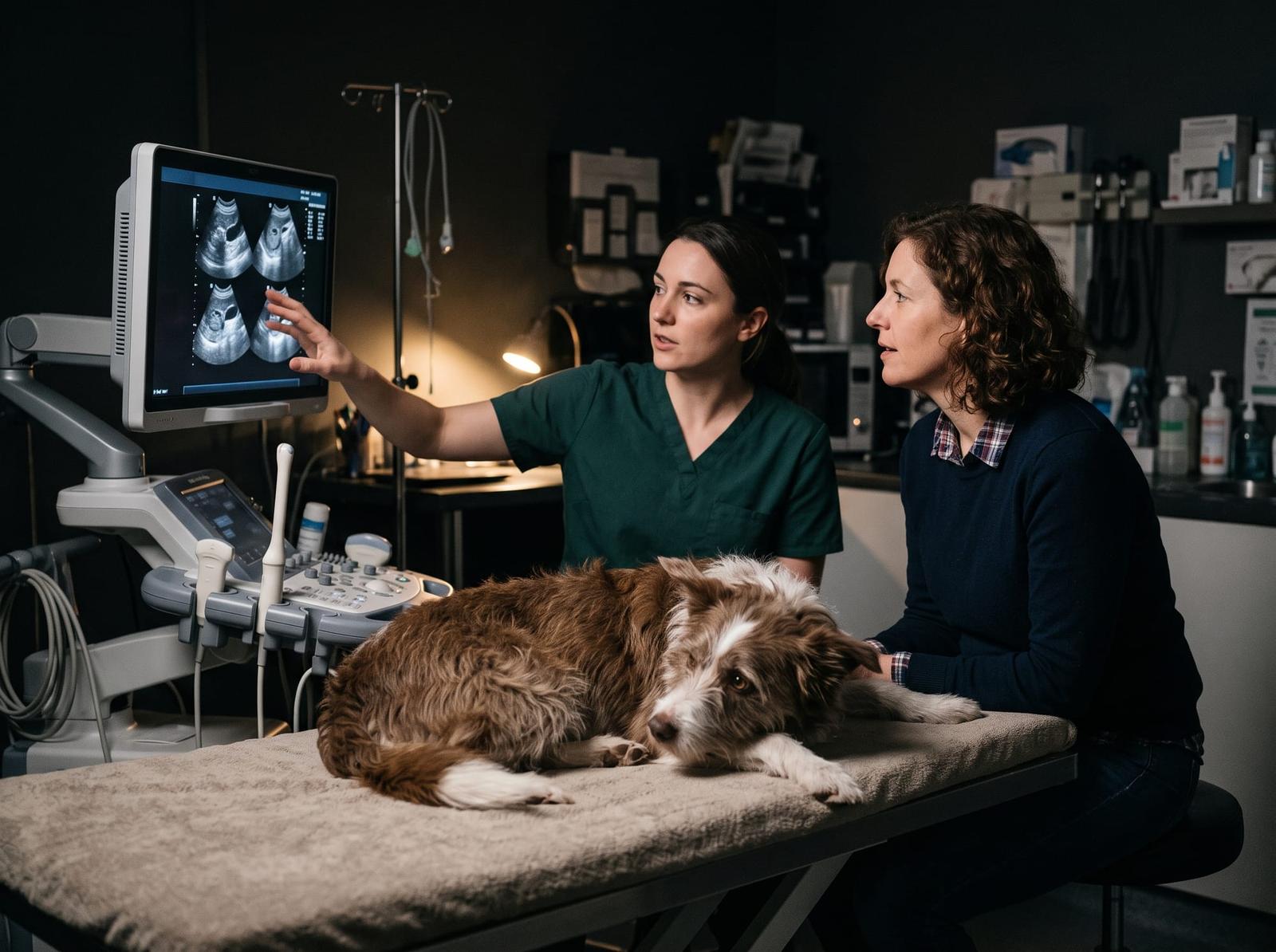
Diagnostic imaging lets us look inside your pet's body safely and without surgery. Each type shows something different: ultrasound shows soft tissues and organs, an echocardiogram shows the heart, and fluoroscopy shows organs moving in real time. CT scans are coming soon.
Every study is interpreted by a board-certified veterinary radiologist, and our team will help you decide which one your pet needs.
Ultrasound
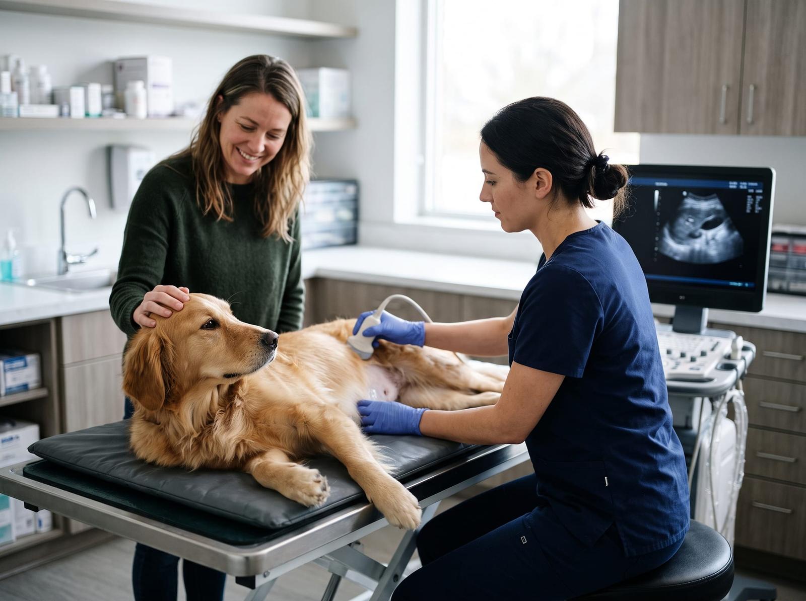
Ultrasound creates real-time images of your pet's internal organs. It is safe and non-invasive, and it is excellent for viewing soft tissues such as the liver, kidneys, spleen and heart, and for checking blood flow. It is often used alongside X-rays.
What we use ultrasound for:
- Abdominal ultrasound: belly pain or swelling; masses, foreign bodies or fluid; changes in the liver, kidneys or pancreas, such as tumors, cysts or inflammation; chronic vomiting, diarrhea or intestinal blockages; bladder stones and urinary tract disease; and checking a pregnancy.
- Neck (cervical) ultrasound: the thyroid, parathyroid and salivary glands, and lymph nodes, for tumor spread and inflammation.
- Ultrasound-guided sampling: safely guiding a needle to collect cells or a tissue sample from a specific organ or mass. A fine needle aspirate uses a very small needle (25G or 22G) with no skin incision. A core needle biopsy uses a slightly larger needle (14G, 16G or 18G) and a small skin incision.
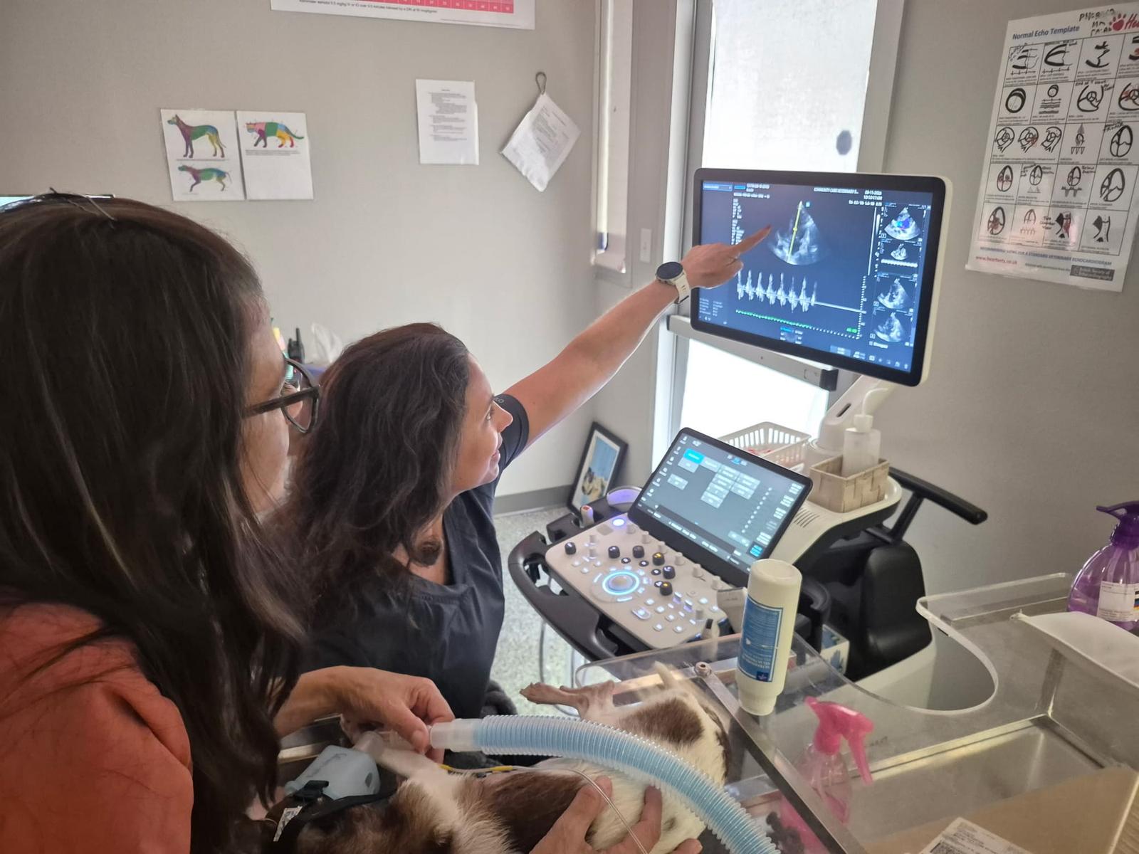
Echocardiograms
An echocardiogram, or echo, is a type of ultrasound used only to look at the heart. It gives a detailed, real-time view of the heart's structure, how well it works and how blood flows through it.
An echo can help:
- Evaluate heart murmurs and find their cause and severity.
- Identify and stage chronic valve disease, such as mitral valve disease, which is common in small-breed dogs.
- Diagnose heart muscle disease, such as dilated cardiomyopathy (DCM) or hypertrophic cardiomyopathy (HCM, common in cats).
- Measure how well the heart pumps and the size of its chambers.
- Find heart defects a pet was born with.
- Check for fluid around the heart (pericardial effusion).
Fluoroscopy
Fluoroscopy is real-time, moving X-ray imaging shown on a monitor. It lets us watch your pet's organs actively working, so it can find functional problems that standard still X-rays often miss completely.
We most often use fluoroscopy to look at:
- Swallowing and digestion: watching how food and liquid move from the mouth through the esophagus, to diagnose megaesophagus, swallowing disorders or motility problems.
- The airway and breathing: finding a collapsing trachea or bronchial collapse by watching the airway while your pet is coughing or breathing heavily.
- Blood flow and minimally invasive procedures: guiding procedures in real time, such as placing stents for urinary tract problems, or checking blood vessel problems like portosystemic shunts.
CT scans
We're adding CT (computed tomography) scans at CCVS Meadowbrook. Check back here for details.
Coming Soon
Reading the images, and getting ready
Why a board-certified radiologist matters
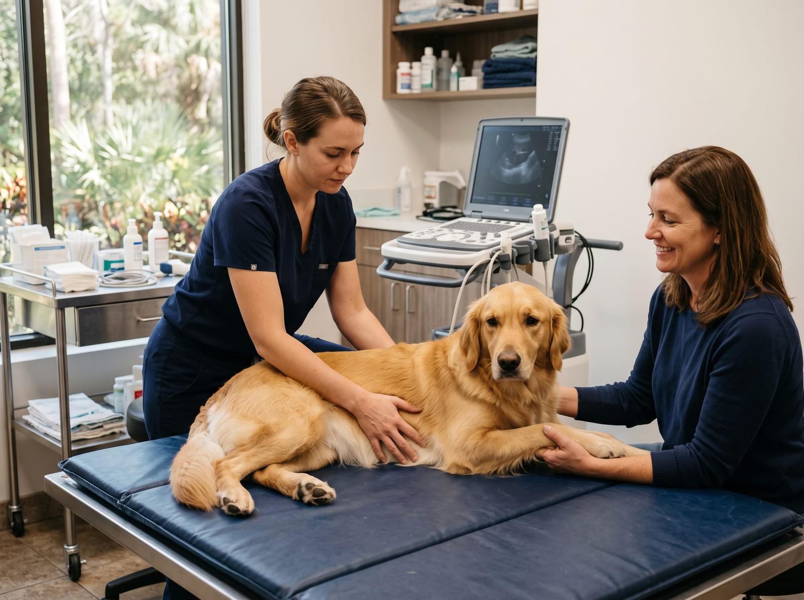
Radiologists have dedicated, intensive training in interpreting diagnostic images, so they can spot subtle problems that may otherwise be missed. That leads to a more accurate and timely diagnosis, which is critical to planning the most effective treatment. Our in-house specialists work directly with your primary veterinarian and our medical team to keep your pet's care seamless.
Preparing your pet
Your pet may be prescribed anti-nausea and/or anti-anxiety medication to keep the visit smooth and comfortable. Fasting may also be recommended. For fluoroscopy this is usually at least 8 hours, especially for a swallow study, a look at the digestive tract, or if mild sedation might be needed.
What to send before scheduling
- Medical history: existing conditions, and a clear timeline of your pet's symptoms.
- Primary veterinary records: medical notes, exam findings and lab work from your primary veterinarian.
- Previous imaging: any earlier X-rays, ultrasounds or echos that can be used for comparison.
- Current medications: an up-to-date list. Before a fluoroscopy study we may ask you to pause some, such as cough suppressants or stomach and gut medications.
Not sure where to start? Call CCVS Meadowbrook.
Our team will help you decide what your pet needs next. In an emergency, come straight in during ER hours: 7am to midnight, 365 days a year.
